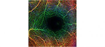
Human Retinal Microvasculature
Detailed visualization of human retinal microvasculature with speckle variance optical coherence tomography (svOCT).
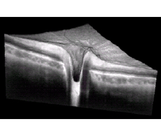
OCT Imaging of Human Retina
Representative images of in vivo human retina/choroid/optic nerve head using 1060-nm swept-source optical coherence tomography (OCT) system.
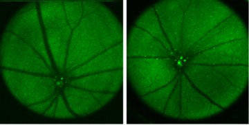
Imaging AD Mouse Retina with fSLO
Images of Alzheimer’s disease model mouse retina were taken using fluorescent scanning laser ophthalmoscope (fSLO) for investigations on Aβ deposits in the eye.
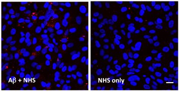
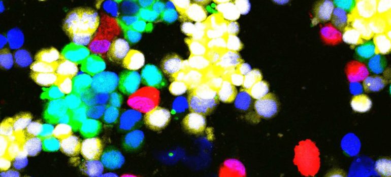
Amyloid beta Induced Membrane Attack Complex (MAC) Formation on RPE Cells
Membrane attack complex (MAC) is formed under the combined stimulation of amyloid beta (Aβ) and normal human serum (NHS), immunolabeled with a monoclonal mouse anti-human C5b-9 antibody and subsequently visualized by Cy3 (red). RPE cell nuclei are counter-stained with DAPI. Scale bars: 20 μm. See full article online.
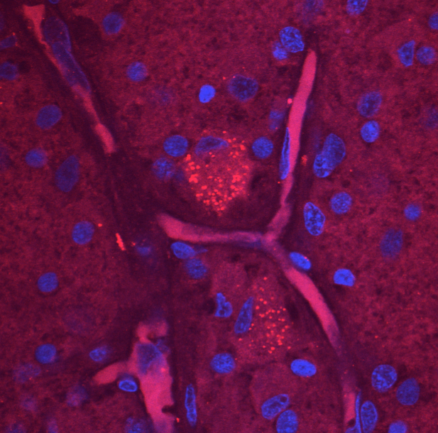
Amyloid Beta and Retinal Ganglion Cells
Confocal image showing ganglion cells (blue nuclei) of a retinal whole mount, immunolabeled with amyloid beta (red puncta).